Sometimes I feel lucky that I was born in an era where technology has brought a revolution to the whole world. And today, technology has made our life so advanced that we can get everything at our doorstep with just a single click of our smartphones. The revolutionary tools, equipments and gadgets have completely changed the way we live our lives as compared to a decade ago. With this advancement, it has become impossible for us to imagine our life without these gadgets and equipment.
But to really appreciate the effects of technology – both its virtues and costs – we need to examine the world of humans before technology. What were our lives like without inventions? For that we need to peek back into the Palaeolithic era when technology was scarce and humans lived primarily surrounded by things they did not make. The advancements in inventions led to discoveries that changed the human lives tremendously. A few noted accidental inventions without which life would have been unimaginably difficult are stated here.
(Pacemaker - The original idea)
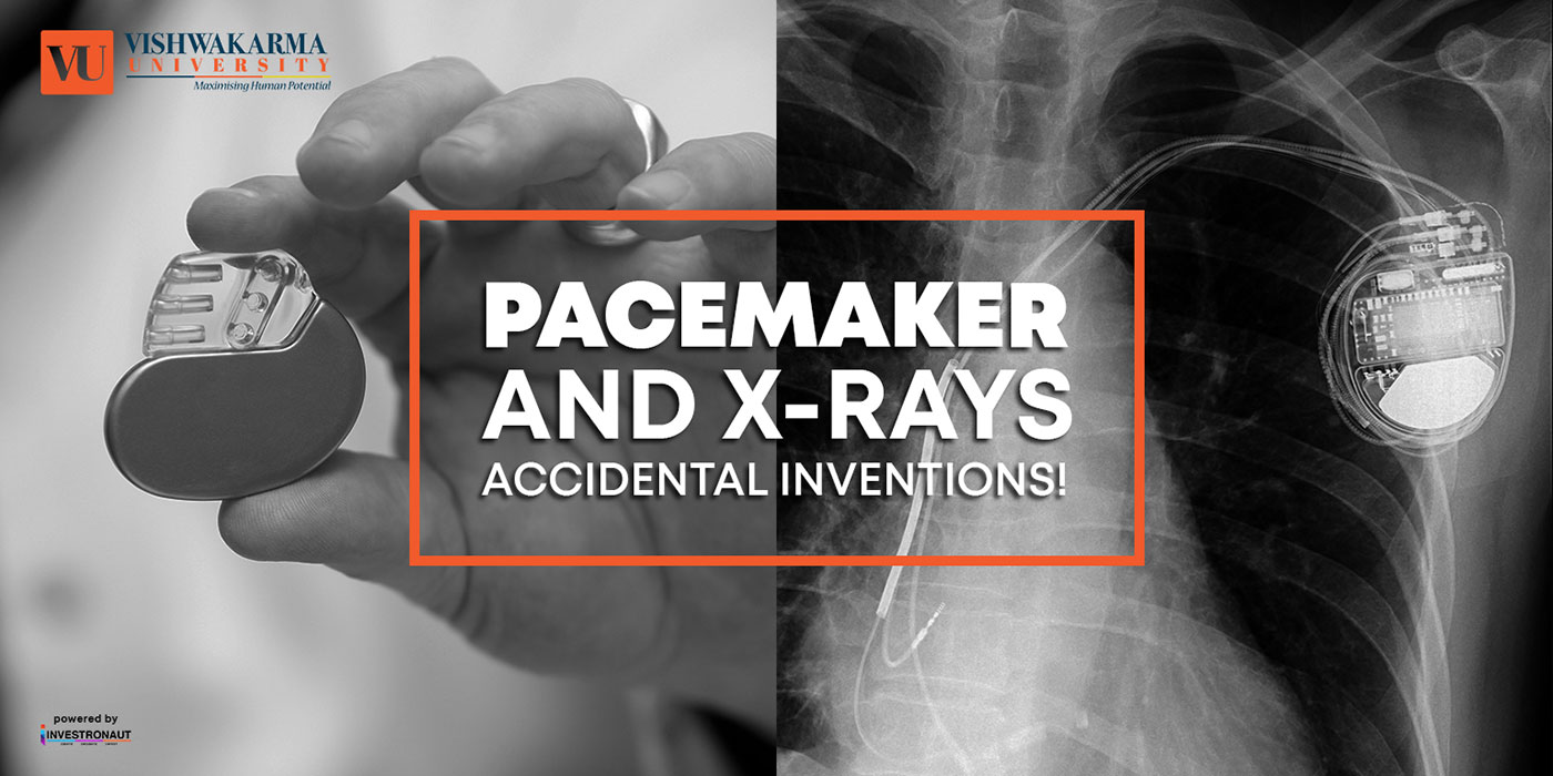
With one of our most vital organs being the heart, conditions such as arrhythmias where the heart either beats too slow, too fast, or with an irregular rhythm can have extremely detrimental effects on one’s everyday life. People may be unable to continue an active lifestyle, suffer breathing problems, and even subsequent organ damage that can lead to terminal ailments or death. A pacemaker helps assuage the problems of arrhythmias to increase longevity and help those with heart conditions to lead a healthier and more active lifestyle. Using electrical pulses, a pacemaker can regulate heartbeats to pump blood throughout the body at a normal rate.
Notable inventor, Wilson Greatbach, invented the first implantable pacemaker by accident while he was attempting to construct an oscillator that would be utilised to record different heart sounds. Pulling one of the resistors from the wrong box led to the advent of the life-saving device that is used prominently today. A rhythmic beating sound was rendered during his flub, and it was then that Greatbach decided to scrap his original invention and create an implantable pacemaker. After two years of fine-tuning the device to perfection, the pacemaker went on to be hailed as “one of the ten greatest achievements of the last 50 years by the National Society of Professional Engineers.”
(WAND - the latest developed pacemaker)
A new Neuro stimulator developed by engineers at the University of California, Berkeley, can listen to and stimulate electric current in the brain at the same time, potentially delivering fine-tuned treatments to patients with diseases like epilepsy and Parkinson’s. The device, named the WAND, works like a "pacemaker for the brain," monitoring the brain's electrical activity and delivering electrical stimulation if it detects something amiss.
These devices can be extremely effective at preventing debilitating tremors or seizures in patients with a variety of neurological conditions. But the electrical signatures that precede a seizure or tremor can be extremely subtle, and the frequency and strength of electrical stimulation required to prevent them is equally touchy. It can take years of small adjustments by doctors before the devices provide optimal treatment.
WAND, which stands for Wireless Artifact -free Neuromodulation Device, is both wireless and autonomous, meaning that once it learns to recognise the signs of tremor or seizure, it can adjust the stimulation parameters on its own to prevent the unwanted movements. And because it is a closed-loop -- meaning it can stimulate and record simultaneously -- it can adjust these parameters in real-time.
(X-rays - The original idea)
If we didn’t have X-rays, would we suddenly just assume we had (or didn’t have) a broken bone? Would surgeons need to merely guess which part of the body is fractured? And what would we be doing during this time when the pain of the broken bone gets unbearable and the doctor is still figuring out which one is broken?I digress…
X-rays are an integral part of the medical field, as they can show medical professionals if and where a broken bone or fracture has occurred, where a bullet is lodged, signs of pneumonia and they are also used to identify breast cancer with mammograms. The use of X-rays has become so standard in medical practice, it is hard to believe that the invention of the X-ray was a complete accident.
In 1895, physicist Wilhelm Conrad Rontgen was spending time in his lab in Germany to try and figure out if cathode rays were able to pass through glass (you know, typical physics stuff). To block a majority of the radiation, Rontgen had set up thick pieces of cardboard around a fluorescent screen, but was in for a surprise when he noticed a strange glowing on the screen penetrating the cardboard barriers every time he switched on the cathode ray.
While others may have decided that level of radiation was terrifying and just scrapped the project, Rontgen investigated the glowing screen and found that the glowing permeated several objects. He even placed his hand in front of the screen only to be welcomed by the sight of the bones in his hands, thus discovering that the ray could penetrate almost anything except for things like bone and lead.
It took years to perfect X-rays, as scientists and doctors didn’t initially realise the harmful effects of radiation, which can cause fatal conditions like skin cancer. Today, X-rays are used widely in medicine and also in airports for extra security measures.
(CERN - the latest developed X-ray)
What if, instead of a black and white X-ray picture, a doctor of a cancer patient had access to colour images identifying the tissues being scanned? This colour X-ray imaging technique could produce clearer and more accurate pictures and help doctors give their patients more accurate diagnoses.
This is now a reality, thanks to a New-Zealand company that scanned, for the first time, a human body using a breakthrough colour medical scanner based on the Medipix3 technology developed at CERN. Father and son scientists Professors Phil and Anthony Butler from Canterbury and Otago Universities spent a decade building and refining their product.
Medipix is a family of read-out chips for particle imaging and detection. The original concept of Medipix is that it works like a camera, detecting and counting each individual particle hitting the pixels when its electronic shutter is open. This enables high-resolution, high-contrast, very reliable images, making it unique for imaging applications in particular in the medical field.
And ohh yes…all this while we thought science was difficult! I always pondered over the unimaginable inventions and cursed the scientists for being so smart! Now I know the secret behind unbelievable scientific inventions - it was purely accidental.
References :


